Visual and macular anatomical changes in central venous occlusion with vitrectomy and radial optic nerve incision in the world's core medical journals. Background: Central venous obstruction (CRVO) is a common retinal vascular abnormality that is the main cause of vision loss. The cause is usually thought to be caused by vascular occlusion in the sieve plate area. This study was designed to evaluate the potential advantages of neuroradiology (RON) and to determine changes in macular thickness and macular volume in CRVO patients. METHODS: Ten patients with CRVO were enrolled in a prospective pilot study. The visual acuity (VA) score was obtained by the Diabetic Retinopathy Early Treatment Study Scale and the corresponding Snellen Vision Examination before and 6 months after surgery. Color fundus photography, fluorescent angiography (FA) and OCT were examined before and 2, 6, 12, and 24 weeks after surgery, and macular thickness and macular volume were evaluated by OCT. RESULTS: Reperfusion was observed in 4 of 10 patients, and 8 visual acuity scores were improved. The median score increased from 11.50 (0-68) to 35.00 (3179). Of the 7 patients, 6 had a reduction in macular volume, a median volume of 77), and 6 had chorioretinal venous traffic, and 6 OCT showed persistent macular edema. CONCLUSIONS: A corresponding decrease in VA score, macular thickness, and macular volume was observed after RON, but 60% of patients developed persistent macular edema. The current selection of the best treatment criteria remains controversial, and this pilot study suggests that RON is beneficial for VA improvement in CRVO patients.
Eyeball contusion secondary to retinal vascular occlusion Purpose: This article describes a rare type of fundus abnormality and its pathological basis after eyeball contusion. METHODS: A prospective, observational case series of 5 consecutive male patients with retinal vascular occlusion after contusion. RESULTS: The results of the study showed that the five patients with retinal vascular occlusion secondary to ocular contusion were found to have lesions by conventional fluorescent fundus angiography, while other subjects were examined for normal subjects. Conclusion: Different forms of retinal vascular occlusion can occur after eyeball contusion. The pathogenesis of this type of occlusion may be related to direct damage to retinal vascular endothelial cells.
Ethyl carbazate Basic Information
Product Name: ethyl carbazate
CAS: 4114-31-2
MF: C3H8N2O2
MW: 104.11
EINECS: 223-903-3
Mol File: 4114-31-2.mol
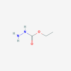
Ethyl carbazate Structure

Melting point 44-47 °C(lit.)
Boiling point 108-110 °C22 mm Hg(lit.)
density 1.2719 (rough estimate)
storage temp. 2-8°C
form Powder
color White to cream
Water Solubility Very soluble in water.
Sensitive Moisture Sensitive
Ethyl Carbazate CAS No. 4114-31-2
ethyl carbazate,ethyl carbazate sds,ethyl carbazate solubility,ethyl carbazate synthesis,Ethyl hydrazinecarboxylate,Hydrazinecarboxylic acid ethyl ester
ShanDong YingLang Chemical Co.,LTD , https://www.sdylhgtrade.com